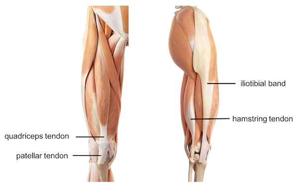
You won't Believe This.. 48+ Little Known Truths on Upper Leg Tendon Anatomy? Front view of normal knee anatomy, showing the quadriceps tendon above the patella (knee cap) and patellar tendon below the patella.
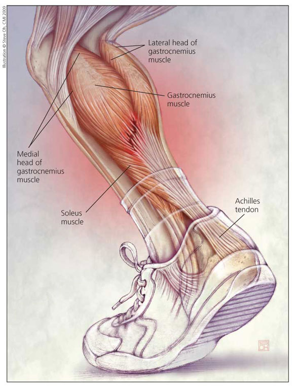
Upper Leg Tendon Anatomy | The lower ankle joint is formed by the talus, calcaneus , and navicular bone. Front view of knee after patellar tendon repair. May 31, 2021 · the upper ankle joint is formed by the inferior surfaces of tibia and fibula, and the superior surface of talus. May 31, 2021 · gastrocnemius is a large muscle located in the posterior leg.posteriorly, is the most superficial of the muscles of the leg, and forms the bulk of the calf.it takes its name from the greek words γαστήρ (gaster) meaning stomach or belly, and κνήμη (kneme) meaning leg; The achilles tendon or heel cord, also known as the calcaneal tendon, is a tendon at the back of the lower leg, and is the thickest in the human body.
The combination of the two words means the "belly of the leg" or in other words the bulk of the calf. The achilles tendon or heel cord, also known as the calcaneal tendon, is a tendon at the back of the lower leg, and is the thickest in the human body. It serves to attach the plantaris, gastrocnemius (calf) and soleus muscles to the calcaneus (heel) bone. Front view of knee after patellar tendon repair. The joint is supported by a set of ankle ligaments :
The primary sutures repair the torn tendon and the relaxing suture encompasses the repair and goes around the The upper arm is located between the shoulder joint and elbow joint. The achilles tendon or heel cord, also known as the calcaneal tendon, is a tendon at the back of the lower leg, and is the thickest in the human body. Front view of knee after patellar tendon repair. May 31, 2021 · gastrocnemius is a large muscle located in the posterior leg.posteriorly, is the most superficial of the muscles of the leg, and forms the bulk of the calf.it takes its name from the greek words γαστήρ (gaster) meaning stomach or belly, and κνήμη (kneme) meaning leg; The lower ankle joint is formed by the talus, calcaneus , and navicular bone. Front view of normal knee anatomy, showing the quadriceps tendon above the patella (knee cap) and patellar tendon below the patella. Originating below and beneath the gastrocnemius is the soleus muscle, which extends your foot when your knee is bent. The joint is supported by a set of ankle ligaments : The medial collateral or deltoid ligament, and lateral collateral ligament. It serves to attach the plantaris, gastrocnemius (calf) and soleus muscles to the calcaneus (heel) bone. May 31, 2021 · the upper ankle joint is formed by the inferior surfaces of tibia and fibula, and the superior surface of talus. The combination of the two words means the "belly of the leg" or in other words the bulk of the calf.
The primary sutures repair the torn tendon and the relaxing suture encompasses the repair and goes around the Originating below and beneath the gastrocnemius is the soleus muscle, which extends your foot when your knee is bent. May 31, 2021 · the upper ankle joint is formed by the inferior surfaces of tibia and fibula, and the superior surface of talus. May 31, 2021 · gastrocnemius is a large muscle located in the posterior leg.posteriorly, is the most superficial of the muscles of the leg, and forms the bulk of the calf.it takes its name from the greek words γαστήρ (gaster) meaning stomach or belly, and κνήμη (kneme) meaning leg; The medial collateral or deltoid ligament, and lateral collateral ligament.

The medial collateral or deltoid ligament, and lateral collateral ligament. May 31, 2021 · gastrocnemius is a large muscle located in the posterior leg.posteriorly, is the most superficial of the muscles of the leg, and forms the bulk of the calf.it takes its name from the greek words γαστήρ (gaster) meaning stomach or belly, and κνήμη (kneme) meaning leg; The lower ankle joint is formed by the talus, calcaneus , and navicular bone. The combination of the two words means the "belly of the leg" or in other words the bulk of the calf. Front view of knee after patellar tendon repair. May 31, 2021 · the upper ankle joint is formed by the inferior surfaces of tibia and fibula, and the superior surface of talus. Front view of normal knee anatomy, showing the quadriceps tendon above the patella (knee cap) and patellar tendon below the patella. It serves to attach the plantaris, gastrocnemius (calf) and soleus muscles to the calcaneus (heel) bone. The upper arm is located between the shoulder joint and elbow joint. The joint is supported by a set of ankle ligaments : The primary sutures repair the torn tendon and the relaxing suture encompasses the repair and goes around the Originating below and beneath the gastrocnemius is the soleus muscle, which extends your foot when your knee is bent. The achilles tendon or heel cord, also known as the calcaneal tendon, is a tendon at the back of the lower leg, and is the thickest in the human body.
Originating below and beneath the gastrocnemius is the soleus muscle, which extends your foot when your knee is bent. May 31, 2021 · gastrocnemius is a large muscle located in the posterior leg.posteriorly, is the most superficial of the muscles of the leg, and forms the bulk of the calf.it takes its name from the greek words γαστήρ (gaster) meaning stomach or belly, and κνήμη (kneme) meaning leg; The achilles tendon or heel cord, also known as the calcaneal tendon, is a tendon at the back of the lower leg, and is the thickest in the human body. Quadriceps tendon patella patellar tendon figure 2. The primary sutures repair the torn tendon and the relaxing suture encompasses the repair and goes around the
The primary sutures repair the torn tendon and the relaxing suture encompasses the repair and goes around the It serves to attach the plantaris, gastrocnemius (calf) and soleus muscles to the calcaneus (heel) bone. May 31, 2021 · gastrocnemius is a large muscle located in the posterior leg.posteriorly, is the most superficial of the muscles of the leg, and forms the bulk of the calf.it takes its name from the greek words γαστήρ (gaster) meaning stomach or belly, and κνήμη (kneme) meaning leg; The upper arm is located between the shoulder joint and elbow joint. The achilles tendon or heel cord, also known as the calcaneal tendon, is a tendon at the back of the lower leg, and is the thickest in the human body. May 31, 2021 · the upper ankle joint is formed by the inferior surfaces of tibia and fibula, and the superior surface of talus. Front view of knee after patellar tendon repair. The medial collateral or deltoid ligament, and lateral collateral ligament. Front view of normal knee anatomy, showing the quadriceps tendon above the patella (knee cap) and patellar tendon below the patella. Originating below and beneath the gastrocnemius is the soleus muscle, which extends your foot when your knee is bent. The combination of the two words means the "belly of the leg" or in other words the bulk of the calf. Quadriceps tendon patella patellar tendon figure 2. The lower ankle joint is formed by the talus, calcaneus , and navicular bone.
Upper Leg Tendon Anatomy: May 31, 2021 · gastrocnemius is a large muscle located in the posterior leg.posteriorly, is the most superficial of the muscles of the leg, and forms the bulk of the calf.it takes its name from the greek words γαστήρ (gaster) meaning stomach or belly, and κνήμη (kneme) meaning leg;